
Modern X‑ray technology allows us to see beyond what is visible during a standard exam. With high‑resolution 3D and panoramic imaging, we can evaluate bone, teeth, joints, sinuses, and soft tissues with exceptional clarity. This ensures accurate diagnosis and safer, more effective treatment planning.
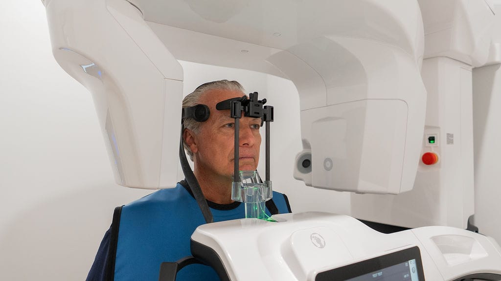
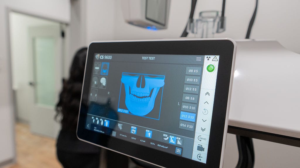
3D cone beam computed tomography (CBCT) provides a complete, three‑dimensional view of your oral and facial structures. This technology:
Captures a full 3D scan in about 20 seconds
Uses significantly less radiation than a medical CT scan
Reveals details that traditional 2D X‑rays cannot
Allows precise planning for implants, extractions, grafting, and more
CBCT imaging is one of the most valuable tools for predictable, high‑quality surgical outcomes.
A panoramic X‑ray provides a broad, two‑dimensional image of your entire mouth in a single scan. It is commonly used to evaluate:
Tooth positioning
Jaw joint (TMJ) health
Sinus conditions
Bone loss from periodontal disease
Abscesses or infections
Developmental abnormalities
Panoramic imaging is fast, comfortable, and essential for diagnosing many oral conditions.


Yes. CBCT uses significantly less radiation than a medical CT scan and is safe for routine diagnostic use.
In most cases, yes. 3D imaging ensures implants are placed safely and accurately.
A full 3D scan typically takes about 20 seconds.
No. Imaging is quick, comfortable, and completely non‑invasive.
Yes. 3D and panoramic imaging can reveal infections, cysts, bone loss, impacted teeth, and other issues not visible during a standard exam.